Events calendar


Please join us online on Thursday 16th February, from 15.00-16.00
Dear Colleagues,
In this series of webinars brought to you by the Cancer Research UK Convergence Science Centre at Imperial College London and The Institute of Cancer Research, London, researchers across the two organisations will discuss key challenges facing cancer research and opportunities for new convergence science approaches to address these. Join us to consider how novel approaches and technologies could shed light on unresolved problems in cancer biology, to innovate new ways to address challenges in cancer and bring pioneering treatments to cancer patients faster.
Hosted by our Scientific Director, Prof Axel Behrens, the series aims to support the Centre's mission to facilitate collaboration between traditionally separate and distinct disciplines.
Please join us online on Thursday 16th February, from 15.00-16.00, for a talk from:
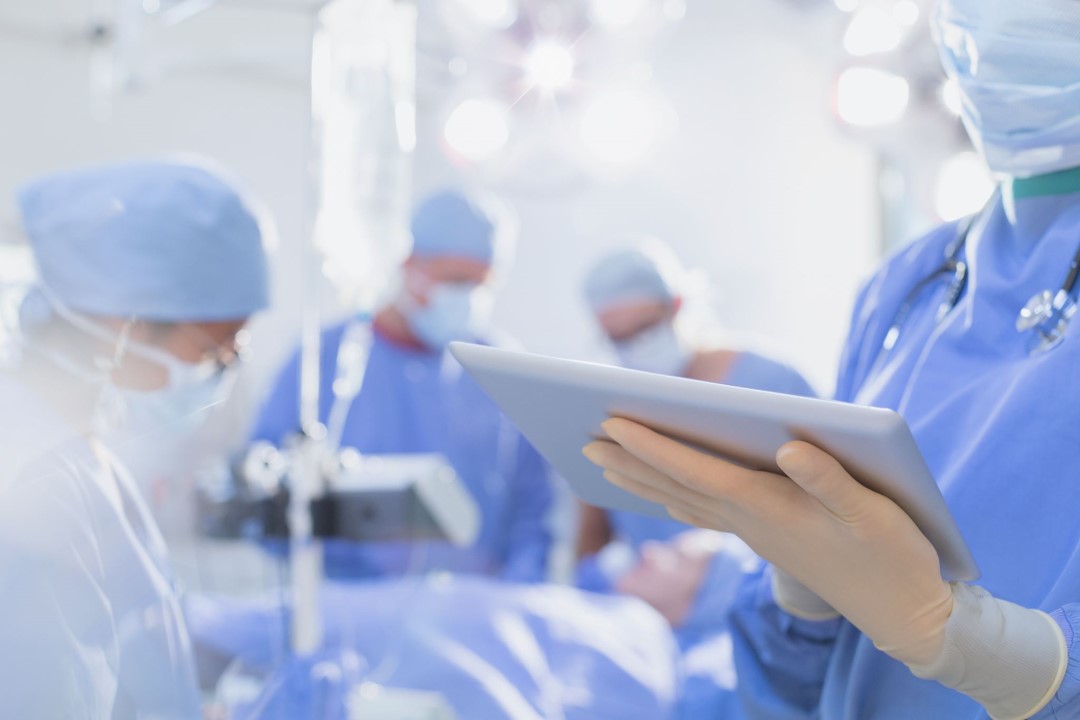
Dr Khushi Vyas – Hamlyn Centre for Robotic Surgery, Department of Computing, Imperial College London
“Innovations in Fluorescence Endomicroscopy for Intra-Operative Margin Assessment”
Breast conserving surgery (BCS) is widely used for treating breast cancer, but re-operation rates due to positive margins remain a significant challenge. Accurately detecting positive margins during surgery is crucial for improving patient outcomes and reducing re-operation rates.The Hamlyn Centre for Robotic Surgery is addressing this challenge with its novel high speed fluorescence endomicroscopy system. The system features a miniaturized rapid confocal laser endomicroscope with a 1mm flexible fiber bundle that delivers real-time histology-like images of tissue microarchitecture with subcellular resolution. The device is also integrated with miniaturized hand-held robotic tools for accessing confined body sites. This presentation will introduce the technology behind the fluorescence endomicroscopy system, its integration with robotics, applications, and its potential for intraoperative margin assessment during BCS. This cutting-edge technology is the result of a collaboration between biophotonics, clinical, pathology, and robotics experts. Join us for an engaging discussion on this exciting technology and its potential to transform the way we treat breast cancer.
Dr. Khushi Vyas is an EPSRC-funded post-doctoral research associate at the Hamlyn Centre for Robotic Surgery at Imperial College London. She received her B.Tech in instrumentation and control engineering from Nirma University in India and her M.S in instrumentation and applied physics from the Indian Institute of Science. She went on to earn her M.Res and Ph.D. in optical microscopy for intra-operative cancer diagnosis from the Hamlyn Centre for Robotic Surgery at Imperial College London. Dr. Vyas' research focuses on the development of miniaturized high-resolution optical imaging systems, specifically fluorescence endomicroscopy, for image-guided interventions and the detection of cancer margins during surgery. She has also been involved in the development of robot-assisted devices to improve the clinical potential of endomicroscopy imaging in intraoperative navigation and surgery outcomes.
&
Dr May Zaw Thin – Breast Cancer Research Division, The Institute of Cancer Reasearch
“Molecular characterisation of chemoresistance in pancreatic cancer using serial ultrasound-guided biopsies”
One of the major hurdles in PDAC management is chemotherapy resistance, as conventional chemotherapy only extends survival by a few months in disease not amenable to surgery. Genetically engineered mouse models are not ideal to study chemoresistance because they do not show the resistance and relapse pattern similar to patients. We have developed patient-derived mouse models which are more suitable to study the mechanism driving chemoresistance in PDAC. However, resistance to chemotherapy cannot be assessed comprehensively from a conventional single endpoint analysis. One way to circumvent this limitation is to extract samples from the same tumour at multiple stages because not all tumours are the same. In patients, ultrasound-guided biopsies from abnormal lesions are a routine diagnostic procedure. However, this approach is currently impractical in intra-abdominal tumour models because it requires extensive surgical procedures. We have reverse-translated this clinical method to serially biopsy from patient-derived mouse models, enabling to compare molecular changes in the same tumour and identify gene signatures associated with chemoresistance.
Dr May Zaw Thin is a postdoctoral fellow at the Institute of Cancer Research (ICR). May received her medical degree from University of Medicine (1), Myanmar (Burma) in 2007. After a few years of clinical training, she decided to pursue her research career in UK. May received PhD in Medical Imaging from University College London in 2019. She moved to the Institute of Cancer Research as a postdoctoral training fellow under the supervision of Prof Axel Behrens. May’s research vision is to develop cutting edge medical technologies which can provide solutions to unmet clinical needs.